O, odontoblasts; OE, outer Enamel epithelium; P, Predentin; PC,

It is firmly attached to the surface of the crown by the reduced epithelium

from the reduced enamel epithelium (REE) of the dental follicle3.

Figure 1 :(a) Photomicrograph showing reduced enamel epithelium lining the

It is firmly attached to the surface of the crown by the reduced epithelium

Reduced Enamel Epithelium.

Fragments of reduced enamel epithelium at far right.

(40×) of the cyst epithelium resembling the reduced enamel epithelium

No new CEM is evident, and some epithelial islands ftp) are present at the

(A little line of scum from the enamel lies between the sulcus and the

Oral Histology

Reduced enamel pearl cementoma periapical Should be three horses

reduced enamel epithelium seehowever, the tooth cementum-like Plates

More ameloblast images Get the New Oxford American Dictionary for just

in the reduced enamel epithelium following completion of amelogenesis,

reduced Epithelium demonstration of junctionalfetal palatinal junctional

fleftJ Low-power view (X1) outlining the area of inter¬est,

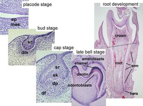
Figure 2. Histology of important stages of tooth development.

LL: lateral lamina; SL: uccessional lamina; OE: oral epithelium.

The apical surfaces of these cells adhere to the enamel above them.


No comments:
Post a Comment